A team of international researchers have created a first-of-its-kind database of ant data, which combines high fidelity 3D scans and genetic information for thousands of specimens covering hundreds of species.
This database, called Antscan, is aimed at enabling new studies of ants by a wider research community, and provides a method for quickly producing similar databases for other animals.
Antscan is a pilot project, led by researchers from the Karlsruhe Institute of Technology in Germany, in collaboration with ANU and the Okinawa Institute of Science and Technology and published in Nature Methods. Their goal is to enhance studies of biodiversity and evolution, by providing rich and up-to-date data sets free to all researchers.
The power of the project is that digital information is accessible from anywhere in the world – for researchers, artists and the public, the research team said.
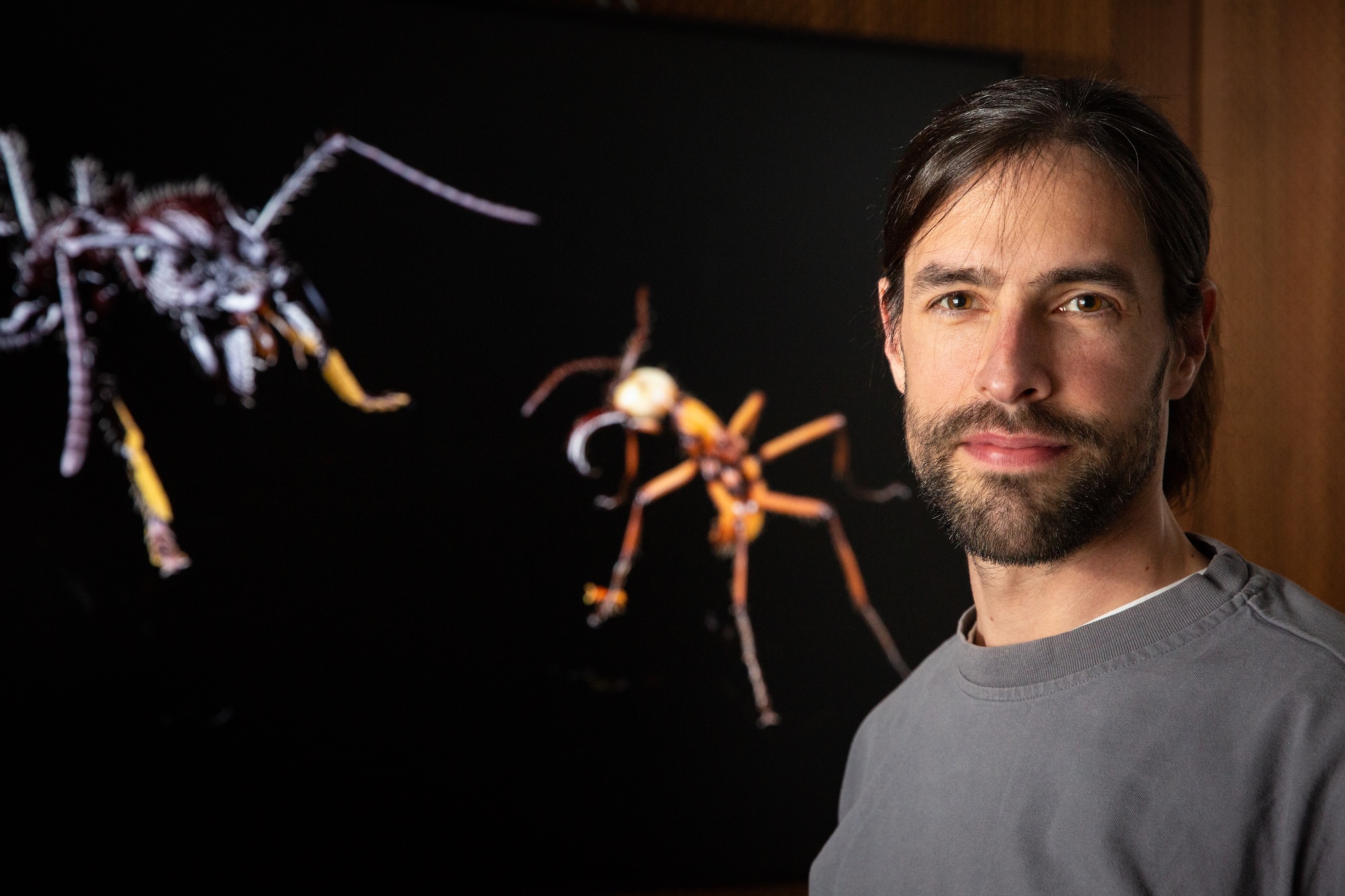
“I want to make the tools and databases I develop accessible to everyone. Seeing other researchers use them for their own discoveries is what really drives me,” said Antscan team member Dr Philipp Loesel, from the ANU Materials Physics Department.
Ants are a prime candidate because they are highly desirable for research on evolution and ecology, with over 14,000 known species spanning ecosystems globally.
The key to creating the ant database is the availability of a new generation of information, beyond photos and surface scans: genetic data and tomography, which can capture the full morphology of an ant, including its internal and external structures.
However, taking advantage of this new information presented a challenge: the time it takes to scan and process x-ray tomography images, which until now could take up to 90 hours to scan and manually post-process for a single specimen.
To overcome this, the Antscan team employed synchrotron micro-tomography, instead of lab-based micro-CT, cutting scan times from ~12 hours to ~2 minutes per scan. Employing this at the Karlsruhe Synchrotron (known as KIT Light Source) enabled the Antscan researchers to scan in thousands of specimens within a single week – a feat that would have taken six years using the previous method.
Once scanned, the specimens need to be segmented. Segmentation requires identifying and separating all parts of a specimen: its exoskeleton, organs and various tissues: typically a laborious process.
To accelerate this process, the researchers called upon Dr Loesel’s Biomedisa package: auto-segmentation software he had previously developed. In work preceding Antscan, the software was capable of reducing the segmentation time of an image of a weevil from 77 hours to just 9 hours.
Initially the team worked on segmenting the ants’ surface, Dr Loesel said.
“We have not automated the segmentation of all parts of the ants. For such a heterogeneous database this is still extremely challenging, and is reserved for future work.”
As images of more ant samples were segmented, Biomedisa improved its efficiency by training AI models on the datasets it had produced. This resulted in a fully automated segmentation process, further reducing post-processing time.
Biomedisa also contributed by providing its website to serve as the portal to access the Antscan database.
The completed Antscan platform is now capable of providing high fidelity, complete, segmented 3D models of roughly 2,200 ant specimens to any researcher connected to the internet. The authors report that to date these cover 792 species, of which 186 species have associated genetic data.
These are just the first steps – Antscan has more room to grow as contributions expand, the authors said.
“With Antscan, we aim to form a platform for future comparative research on the genomic basis of phenotypic variation and diversification in invertebrates,” they said.
The new methodology can easily be applied to other organisms beyond ants - the researchers hope it will be adapted to create similar databases for all kinds of life.
“Antscan establishes a design that can be adapted for other lineages of small organisms across the tree of life, and serves as a vast open resource for promoting research on the morphology and anatomy of ants,” the authors said.
Story: Matthew Wooding


